


Histological Analysis (Histology)
Histology is the scientific study of tissues at a microscopic level. In the context of plastic and reconstructive surgery, histological analysis is a critical diagnostic step following the removal of any skin lesion, tumor, or cyst.
Histological Analysis (Histology)
Histology is the scientific study of tissues at a microscopic level. In the context of plastic and reconstructive surgery, histological analysis is a critical diagnostic step following the removal of any skin lesion, tumor, or cyst. By examining the cellular structure of the removed tissue, a pathologist can provide a definitive diagnosis, confirming whether a growth is benign or malignant and ensuring that the surgical margins are clear of disease.
While a surgeon can often form a clinical suspicion based on the appearance of a growth, the "gold standard" for diagnosis is histology. Every tissue sample removed during surgery is treated as a vital piece of medical evidence. Histology allows us to look beyond what is visible to the naked eye, identifying the specific cell types and growth patterns that define the nature of the lesion.
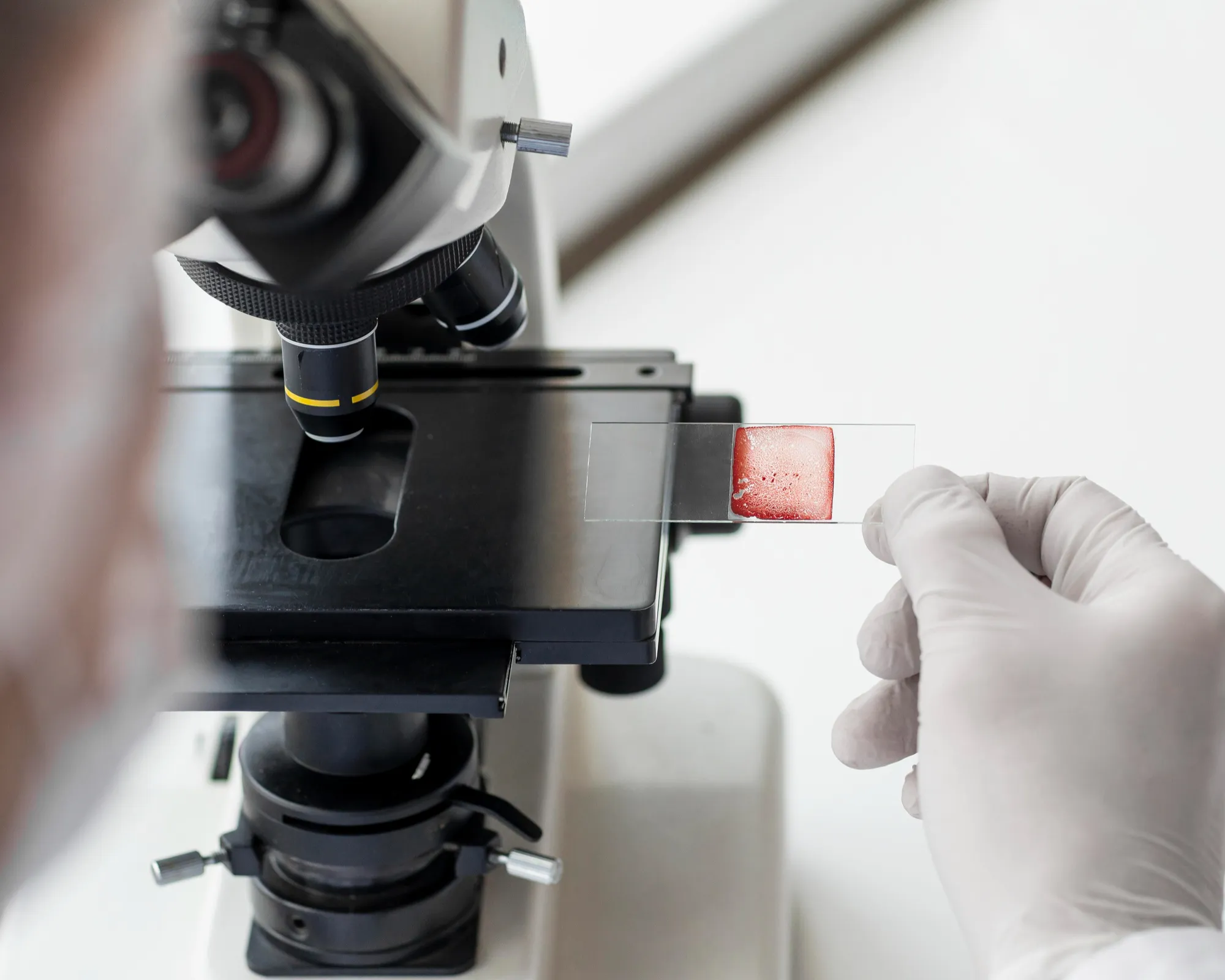
What is Histological Analysis?
The process begins the moment the tissue is removed in the operating room and follows a rigorous path to ensure accuracy:
- Fixation: The tissue specimen is placed in a preservative solution, typically formaldehyde, to prevent decay and maintain the cellular structure in a life-like state.
- Processing and Embedding: The tissue is treated with various chemicals and then embedded in a block of paraffin wax. This provides the structural support necessary to cut the tissue into incredibly thin slices.
- Microtomy: Using a precision instrument called a microtome, the wax block is sliced into sections so thin (often only a few micrometers) that they are translucent. These sections are placed onto glass slides.
- Staining: To make the clear cells visible under a microscope, the slides are treated with specialized dyes. The most common is Hematoxylin and Eosin (H&E), which stains cell nuclei blue and other structures pink, allowing the pathologist to distinguish between healthy and abnormal cells.
- Microscopic Examination: A specialist physician, known as a Pathologist, examines the slides under high magnification to determine the exact diagnosis and check the surgical margins.
Why is histology necessary?
Histology serves several essential functions in your surgical care:
- Definitively identifies the lesion: It distinguishes between common growths like seborrheic keratoses and more serious conditions like melanoma or basal cell carcinoma.
- Confirms "Clear Margins": It verifies that the surgeon has removed a sufficient "safety buffer" of healthy tissue around a tumor, significantly reducing the risk of recurrence.
- Determines the Stage of Disease: For malignant tumors, histology provides details on how deep the cancer has invaded (Breslow depth) and how quickly the cells are dividing (mitotic rate).
- Guides Future Treatment: The results of the histology report dictate whether further surgery, specialized monitoring, or referral to an oncologist is required.
Once the analysis is complete, a formal pathology report is generated. This document typically includes:
- Gross Description: A physical description of the tissue as seen by the naked eye (size, color, and weight).
- Microscopic Description: A detailed account of what the cells look like under the microscope.
- Diagnosis: The final, definitive name of the condition.
- Comments: Additional information regarding the completeness of the excision (the margins) and any specific characteristics of the cells.
Waiting for Results
The process of fixing, embedding, and staining tissue takes time. While some specialized "frozen section" results can be obtained during surgery, a standard formal histology report typically takes between 3 to 7 working days. Your surgeon will review this report in detail and discuss the findings with you during your follow-up appointment.
Is every mole removed sent for histology?
Yes. It is standard surgical practice to send every piece of tissue removed from a patient to the pathology lab. Even if a lesion looks perfectly "normal" to the surgeon, histological analysis is the only way to be 100% certain of the diagnosis and ensure your safety.
What does "Positive Margins" mean?
If a report indicates "positive margins," it means that the pathologist found abnormal cells at the very edge of the removed tissue. This suggests that some of the growth may still remain in the skin. In such cases, your surgeon will usually recommend a "re-excision" to remove a small additional amount of tissue and ensure the area is completely clear.
Are the results ever wrong?
Histology is highly accurate, but like any medical test, it depends on the quality of the sample and the expertise of the pathologist. In rare or complex cases, a second pathologist may be consulted for a "second opinion" to confirm the diagnosis.
Does a histology report mean I have cancer?
Not necessarily. The vast majority of histology reports for routine removals (like cysts, lipomas, or common moles) confirm that the growth is benign (non-cancerous). The report is simply a tool to provide certainty, regardless of the diagnosis.
Every question gets an answer
Get a free consultation by booking a consultation with our consultants.